
Corroyer-Dulmont A, Valable S, Fantin J, Chatre L, Toutain J, Teulier S, Bazille C, Letissier E, Levallet J, Divoux D, Ibazizène M, Guillouet S, Perrio C, Barré L, Serres S, Sibson NR, Chapon F, Levallet G, Bernaudin M
Multimodal evaluation of hypoxia in brain metastases of lung cancer and interest of hypoxia image-guided radiotherapy
Sci. Rep. 2021 11(1):11239
Treatment for patients with brain metastases (BM) remains limited, because not adapted to the microenvironment. Corroyer-Dulmont et al., report at clinical and preclinical levels that hypoxia is a hallmark of the microenvironment of BM from NSCLC lung cancer. BM heterogeneity in terms of hypoxia between patients, and metastases, highlights the potential for personalizing external radiotherapy (RT) to hypoxia. In this study, Hypoxia-image-guided-RT showed a significant interest in comparison to unfocused-brain-RT characterized by an early decrease in tumor cell proliferation and in tumor volume resulting in increased survival.
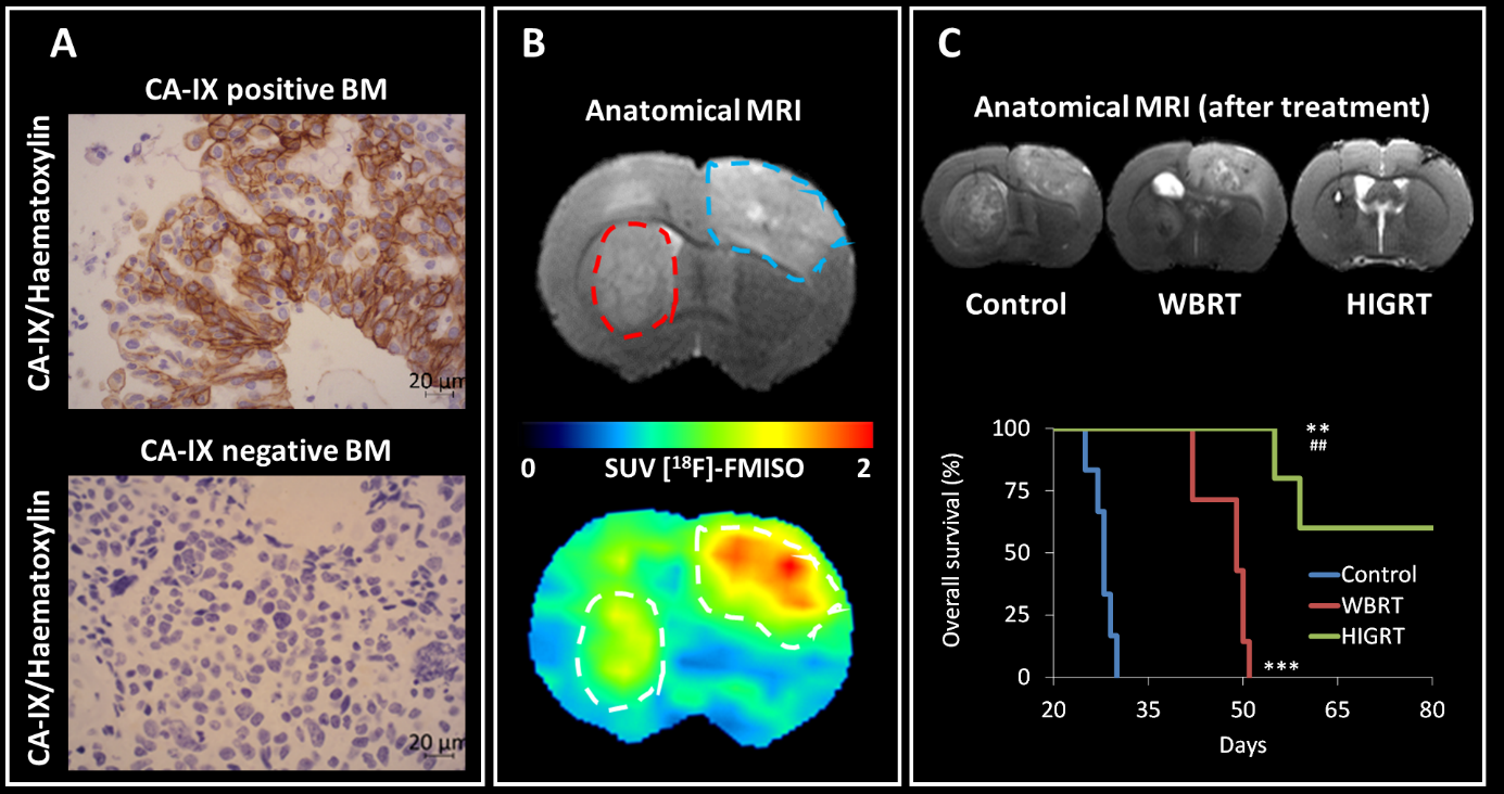
(A) Representative positive and negative CA-IX immunostaining on human brain metastases biopsies. (B) Representative T2w MRI images (top) of rat with cortical (blue hatched line) and striatal (red hatched line) brain metastases in preclinical model. Representative FMISO-PET images (bottom) showing the heterogeneity in terms of hypoxia between brain metastases. (C) Representative T2w MRI images (top) of brain metastases without treatment (control), whole brain radiotherapy (WBRT) and with hypoxia image-guided radiotherapy (HIGRT) showing the therapeutic interest of the approach (bottom), Figure with Open Access Licence – CC BY.
 Français (France)
  English (United Kingdom)